Human Stomach Section Model with Ulcers
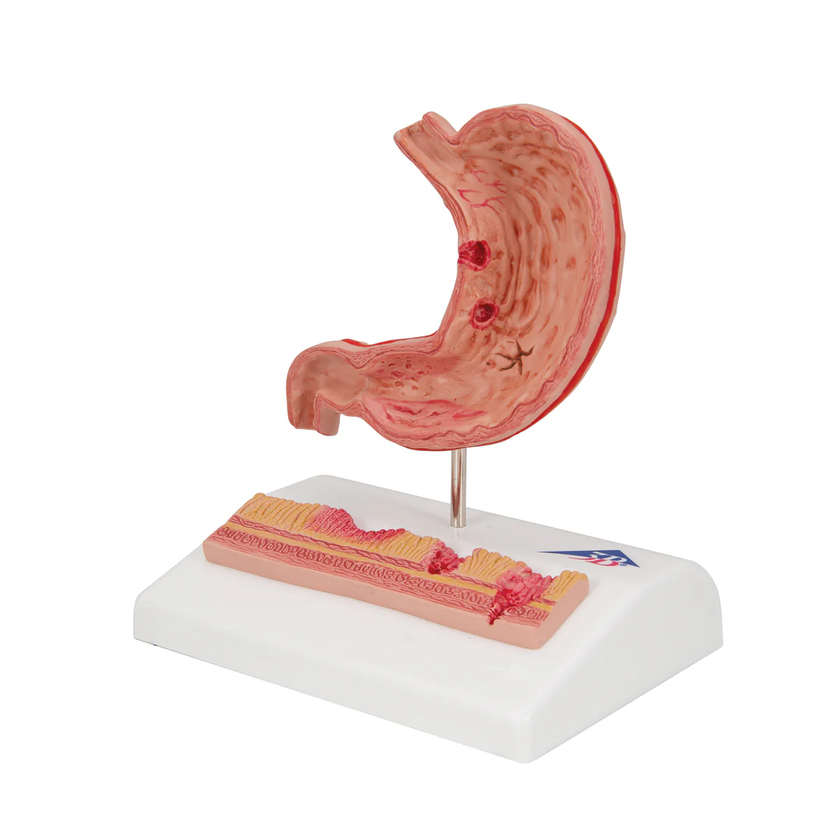
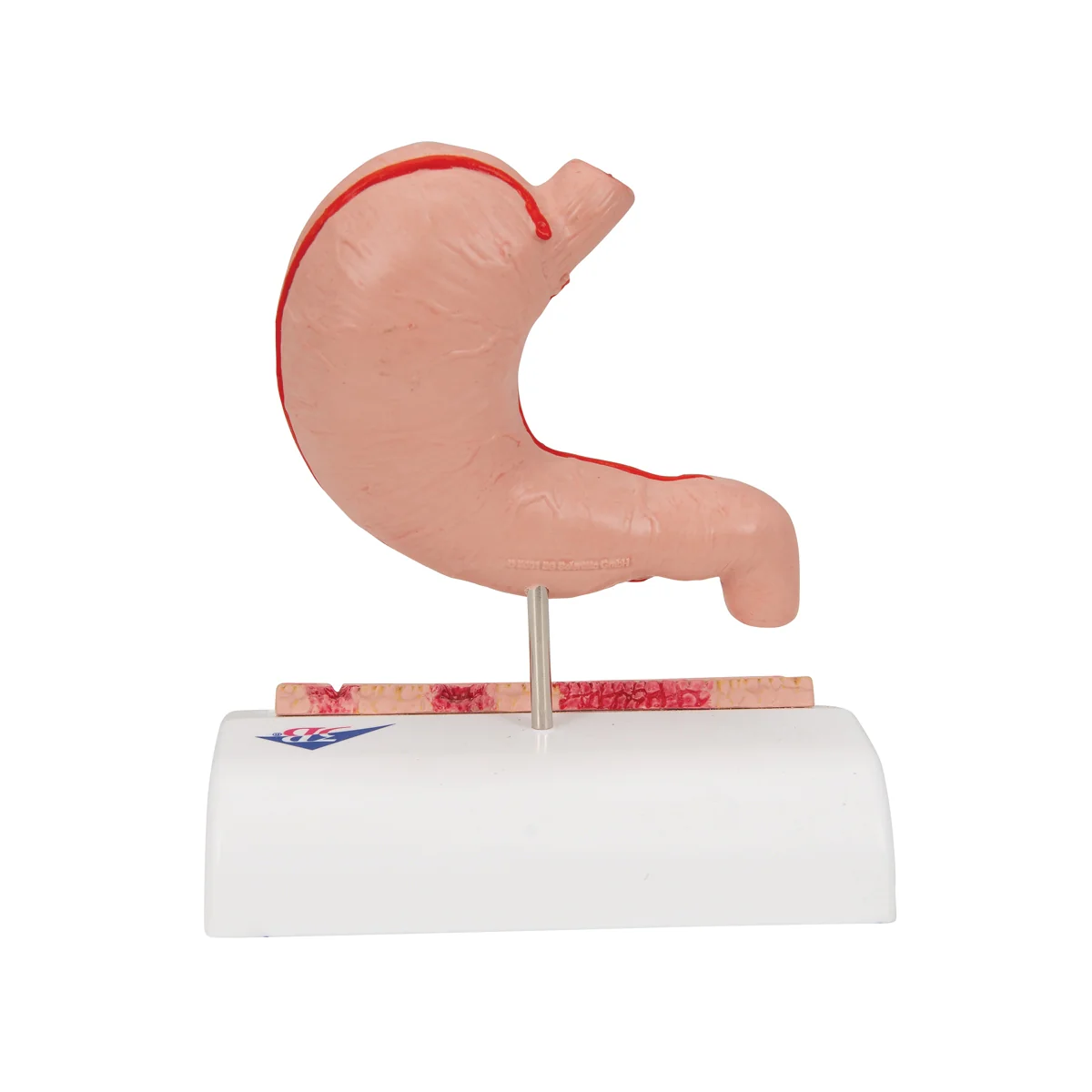
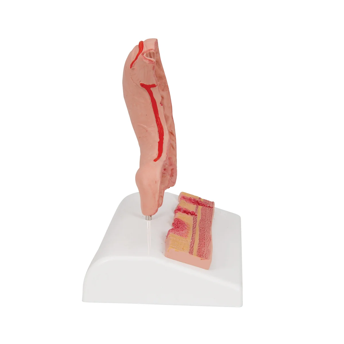
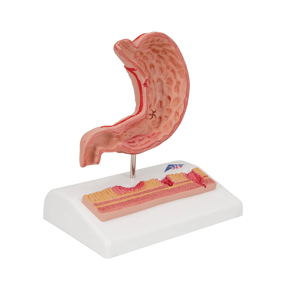
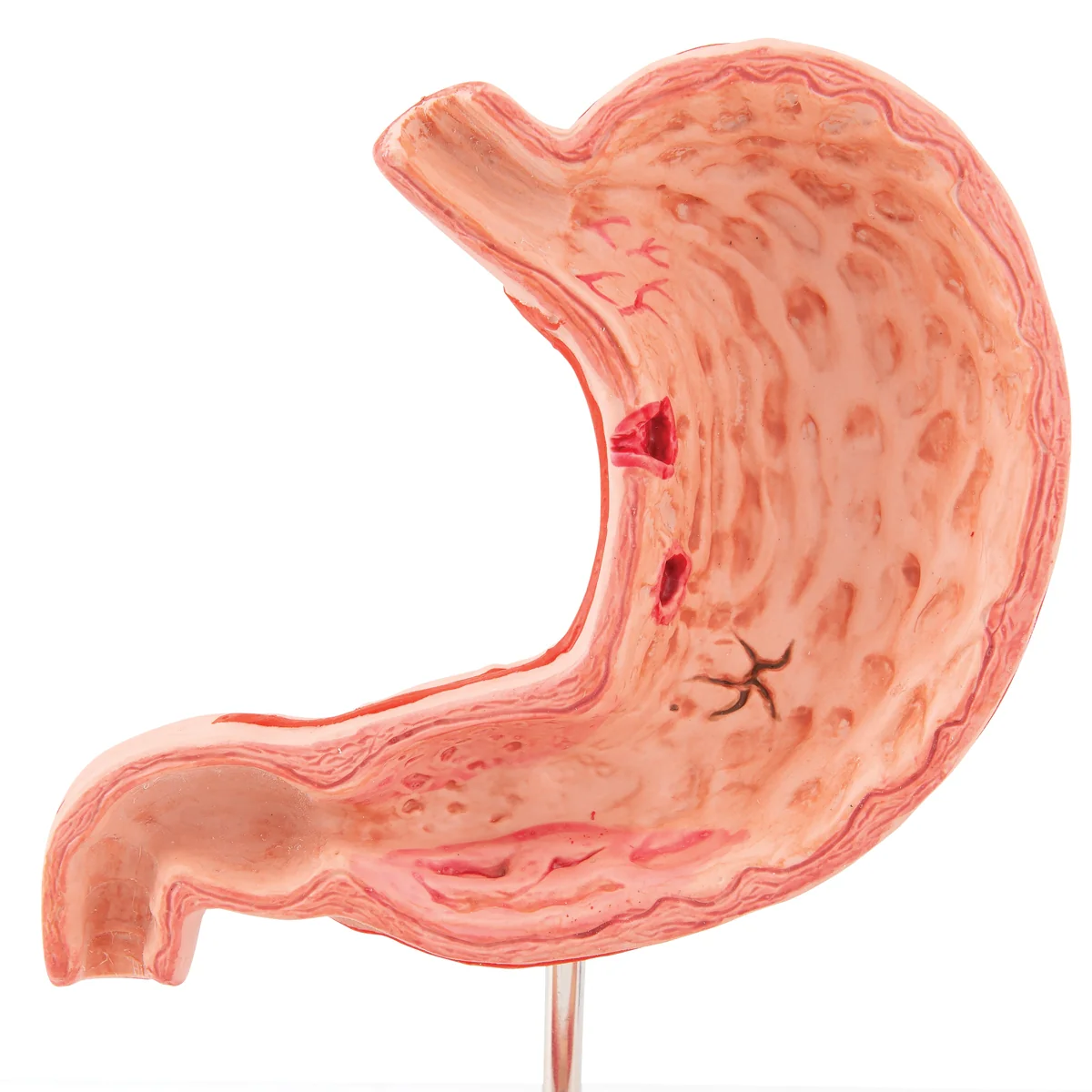
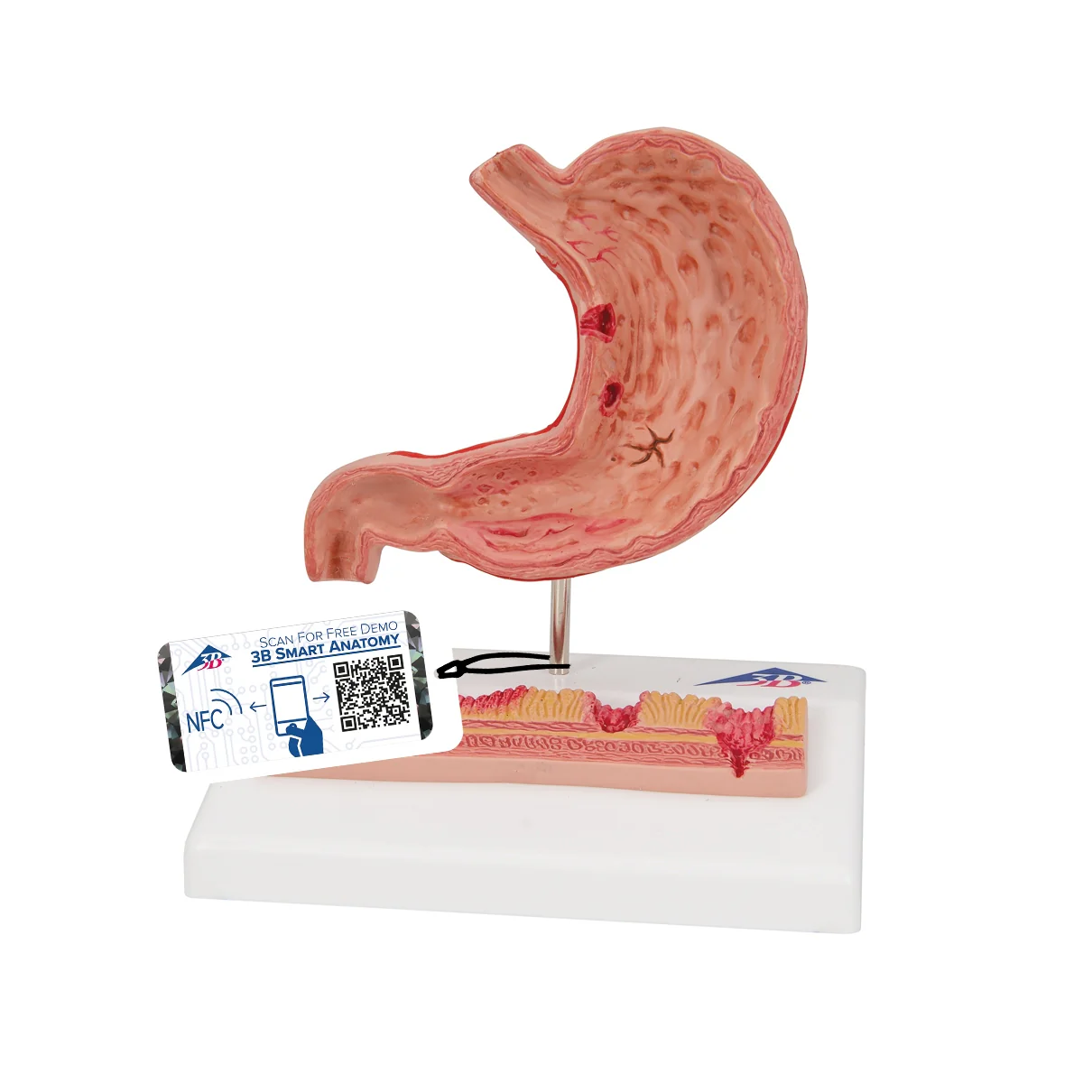
This 3B Scientific® stomach pathology model demonstrates various stages of gastritis from a light gastric ulcer to a perforation. The stomach section with esophagus and duodenum attachment in half life size shows the following pathological changes of the stomach:
- Erythematous gastritis
- Erosive gastritis
- Hemorrhagic gastritis
- Healing stage with scar formation
- Atrophic gastritis
- Hypertrophic gastritis
- Bleeding ulcer
- Perforated ulcer
An additional relief model of the enlarged stomach wall shows:
- Healthy mucous membrane
- Acute gastritis in the antral area
- Erosive gastritis with mucous membrane defects
- Bleeding ulcer (eroded muscularis mucosae)
- Perforated ulcer (all stomach layers eroded)
Stomach with ulcers mounted on a base for display in the classroom or doctors office.
Original: $75.06
-65%$75.06
$26.27More Images

Human Stomach Section Model with Ulcers
This 3B Scientific® stomach pathology model demonstrates various stages of gastritis from a light gastric ulcer to a perforation. The stomach section with esophagus and duodenum attachment in half life size shows the following pathological changes of the stomach:
- Erythematous gastritis
- Erosive gastritis
- Hemorrhagic gastritis
- Healing stage with scar formation
- Atrophic gastritis
- Hypertrophic gastritis
- Bleeding ulcer
- Perforated ulcer
An additional relief model of the enlarged stomach wall shows:
- Healthy mucous membrane
- Acute gastritis in the antral area
- Erosive gastritis with mucous membrane defects
- Bleeding ulcer (eroded muscularis mucosae)
- Perforated ulcer (all stomach layers eroded)
Stomach with ulcers mounted on a base for display in the classroom or doctors office.
Product Information
Product Information
Shipping & Returns
Shipping & Returns
Description
This 3B Scientific® stomach pathology model demonstrates various stages of gastritis from a light gastric ulcer to a perforation. The stomach section with esophagus and duodenum attachment in half life size shows the following pathological changes of the stomach:
- Erythematous gastritis
- Erosive gastritis
- Hemorrhagic gastritis
- Healing stage with scar formation
- Atrophic gastritis
- Hypertrophic gastritis
- Bleeding ulcer
- Perforated ulcer
An additional relief model of the enlarged stomach wall shows:
- Healthy mucous membrane
- Acute gastritis in the antral area
- Erosive gastritis with mucous membrane defects
- Bleeding ulcer (eroded muscularis mucosae)
- Perforated ulcer (all stomach layers eroded)
Stomach with ulcers mounted on a base for display in the classroom or doctors office.